


Gjør som tusenvis av andre bokelskere
Abonner på vårt nyhetsbrev og få rabatter og inspirasjon til din neste leseopplevelse.
Ved å abonnere godtar du vår personvernerklæring.Du kan når som helst melde deg av våre nyhetsbrev.
Develop the strong reasoning skills you'll need for competent and caring practice with An Introduction to Theory and Reasoning in Nursing. This proven book will help you understand theory, what it is, how it supports nurses and their practice and how you can use it to answer clinical questions and care for patients more effectively. -See theorists and their theories come to life with Theorist boxes that contain a brief synopsis of the theorist's life and a photo of the theorist (when available). -Access an alternative to nursing process care plans with a new research-validated reasoning model, the Clinical Reasoning Plan (CRP) that uses actual nursing case studies. -Increase your understanding of how theory and reasoning is used in clinical practice through Nursing Story boxes . -Clarify your understanding of individual support theories and nursing theories through Summaries that identify phenomenon, internal concepts or variables, propositions, external variables, and assumptions. -Increase your mastery of theory, research, and reasoning and their application to nursing practice through Chapter Overviews , Key Words in bold face type, Chapter Outlines. Chapter Introductions, Chapter Summaries, Learning Activities , and a comprehensive Glossary. -Go online to explore specific topics of interest through a wide range of up-to-date References . -Find the information you need fast with anytime, anywhere access to the fully searchable text online.
Covers information on vascular territories, film subtraction, and magnetic resonance angiography. This text is illustrated with 1,200 radiographs and line drawings, and uses boxed summaries to highlight key points.
Epidemiology is the simplest and most direct method of studying the causes of disease in humans, and many major contributions have been made by studies that have demanded nothing more than the ability to count, to think logically, and to have an imaginative idea. With the accumulation of knowledge, however, its has become harder for individuals working alone to make effective contributions, and epidemiological research is becoming increasingly a matter of teamwork, not only because of the large number of people that may have to be studied and the large amount of data that have to be collected and analyzed, but also because of the need to bring together for the design and conduct of the study clinical experience, biological understanding, statistical expertise, and many other special skills that vary from one study to another. But if, in this sense, epidemiological research is becoming more complex, the core of the subject remains essentially simple, and a good epidemiological study should be capable of description in such a way that all who are interested in the cause of disease can follow the argument and decide for themselves the validity of the conclusions.
Features the techniques and electrode placements for common nerve conduction studies. This book describes each nerve conduction study, including placement of electrodes, typical electromyography equipment settings, normal values, and pearls and pitfalls. It provides coverage of surface anatomy for needle electromyography.
Presents strategies for handling a variety of difficult patient interviews. This guide contains chapters which present a hypothetical scenario, describe communication techniques for the phases of the interaction, and identify pitfalls to avoid. It includes examples of physician-patient dialogue, illustrations showing body language, and references.
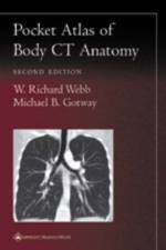
Featuring 229 images, this atlas is a guide to interpreting computed tomography body images. It shows readers to recognize normal anatomic structures on CT scans and distinguish these structures from artifacts. Chapters in this book cover the neck and larynx, thorax, portal venous phase abdomen, pelvis, arterial phase abdomen, and reconstructions.
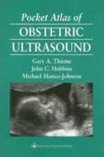
This handy pocket atlas is a complete and convenient guide to the normal sonographic appearances of the embryo and fetus and its uterine environment. The book equips practitioners with the thorough knowledge of normal fetal anatomy that is essential for the timely recognition and diagnosis of abnormalities.
Provides the fundamentals of evaluation and examination techniques of the musculoskeletal system. Each region begins with step-by-step instructions for goniometry, manual muscle testing, muscle length, joint accessory motions and special orthopedic tests. This title includes special discussions of posture and gait analysis.
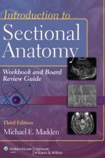
Helps you understand the principles of sectional anatomy. This book contains a variety of exercises and tools that make it easy for you to remember essential information and build critical-thinking skills. It is designed to prepare you for the CT and MR registry examinations.
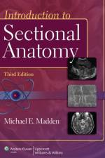
Looking for resource to help your students fully understand and retain critical knowledge of sectional anatomy? This book begins with key terminology and concepts and then moves through the body from head to toe. It includes imaging modalities, ultrasound, MR, and PET/CT, so your students are fully equipped for what they can encounter in practice.
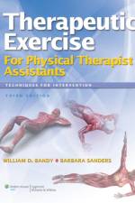
Offers the training you need to fulfill your responsibilities as a physical therapist assistant. This book gives you the knowledge and skills to effectively implement patient treatment plans using therapeutic exercise techniques that you administer under the direction of a physical therapist.
Helps you develop the deep understanding of common patient presentations necessary to prevent diagnostic and treatment errors and to improve outcomes. This book combines evidence-based practice with well-earned experience and best practices opinion to help you avoid common errors of prehospital care.
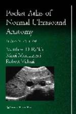
Featuring 74 images, this pocket atlas helps readers identify key anatomic structures of the neck, abdomen, female pelvis, and male genitalia on ultrasound scans and shows how to distinguish these structures from artifacts.
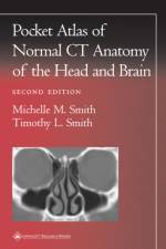
Featuring 73 images, this pocket atlas helps interpret computed tomography images of the brain and calvarium, temporal bone, orbit, nasal cavity, and paranasal sinuses. It also helps readers recognize normal anatomic structures on CT scans and distinguish these structures from artifacts.
Abonner på vårt nyhetsbrev og få rabatter og inspirasjon til din neste leseopplevelse.
Ved å abonnere godtar du vår personvernerklæring.